Research article Materials
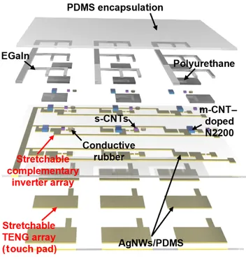
Stretchable Complementary Integrated Electronics Based on Elastic Dual-Type Transistors
Y. Zhang , K. Sim , H. Shim , F. Ershad , S. Patel , Y.-S. Guan , C. Yu
Science Advances · 11(45), eaea1867 (2025)

Showing all 28 papers
University of Houston
Y. Zhang , K. Sim , H. Shim , F. Ershad , S. Patel , Y.-S. Guan , C. Yu
Science Advances · 11(45), eaea1867 (2025)
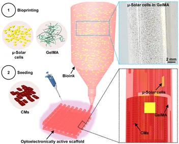
F. Ershad† , Z. Rao† , S. Maharajan† , F. C. Paccola Mesquita , J. Ha , L. Gonzalez , T. Haideri , E. Curty da Costa , A. Moctezuma-Ramirez , Y. Wang , S. Jang , Y. Lu , S. Patel , X. Wang , Y. Tao , J. Weygant , C. Garciamendez-Mijares , L. C. Orrantia Clark , M. Zubair , X. Lian , A. Elgalad , J. Yang , C. Hochman-Mendez , Y. S. Zhang , C. Yu
† Equal contribution
Science Advances · 11(4), eadt7210 (2025)
Prior to University of Houston faculty appointment
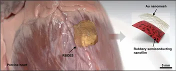
Z. Rao , F. Ershad , Y.-S. Guan , F. C. Paccola Mesquita , E. Curty da Costa , M. A. Morales-Garza , A. Moctezuma-Ramirez , B. Kan , Y. Lu , S. Patel , H. Shim , K. Chen , W. Wu , T. Haideri , X. Lian , A. Karim , J. Yang , A. Elgalad , C. Hochman-Mendez , C. Yu
Science Advances · 10(49), eadq5061 (2024)
Prior to University of Houston faculty appointment
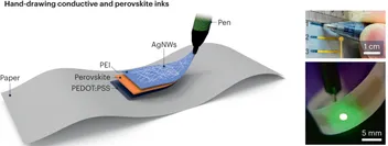
F. Ershad , W. Song , C. Yu
Nature Photonics · 17(11), 928–930 (2023)
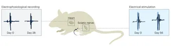
Y. Liu , F. Ershad , Y. Tao , C. Yu
Nature Materials · 22(7), 801–802 (2023)
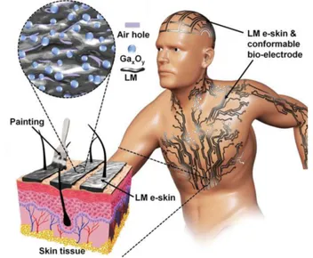
F. Ershad , S. Patel , C. Yu
npj Flexible Electronics · 7(1), 32 (2023)
K. Pu , Z. Xu , Y. Gao , C. Zhao , F. Ershad , Y. Li , R. Wang , C. Yu , G. Wei
Advanced Materials Technologies · 8(16), 2300207 (2023)
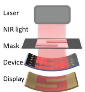
F. Ershad , M. Houston , S. Patel , L. Contreras , B. Koirala , Y. Lu , Z. Rao , Y. Liu , N. Dias , A. Haces-Garcia , W. Zhu , Y. Zhang , C. Yu
PNAS Nexus · 2(1), pgac291 (2023)
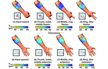
Prior to University of Houston faculty appointment
Y.-S. Guan , F. Ershad , Z. Rao , Z. Ke , E. Curty da Costa , Q. Xiang , Y. Lu , X. Wang , J. Mei , P. Vanderslice , C. Hochman-Mendez , C. Yu
Nature Electronics · 5(12), 881–892 (2022)
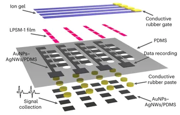
N. Prodan , F. Ershad , A. R. Alcaraz , L. Li , B. Mistretta , L. Gonzalez , Z. Rao , C. Yu , P. Gunaratne , N. Li , R. J. Schwartz , B. K. McConnell
iScience · 25(11), 105402 (2022)
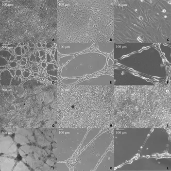
H. Shim , F. Ershad , S. Patel , Y. Zhang , B. Wang , Z. Chen , T. Marks , A. Facchetti , C. Yu
Nature Electronics · 5(10), 660–671 (2022)
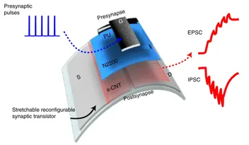
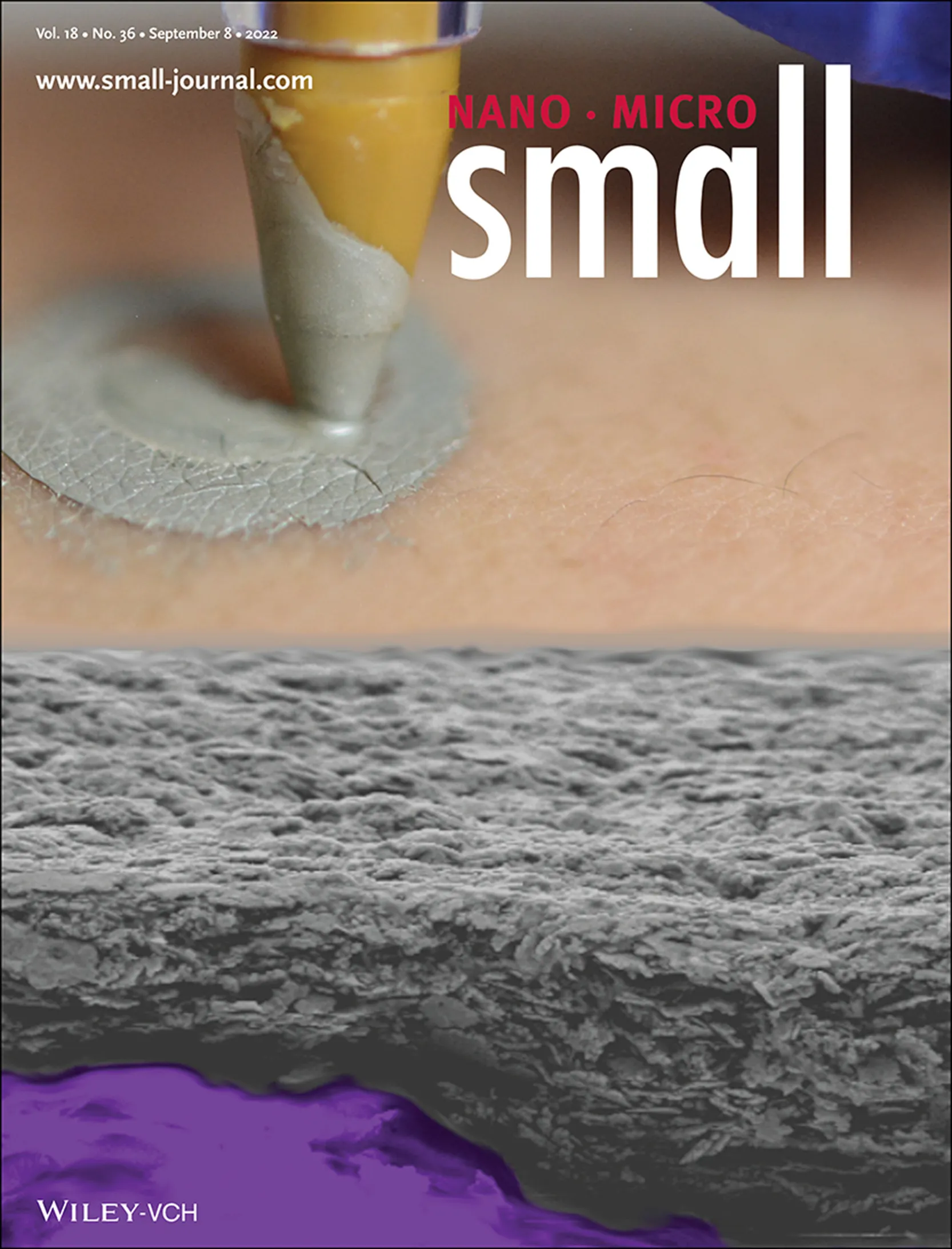
S. Patel† , F. Ershad† , J. Lee , L. Chacon-Alberty , Y. Wang , M. A. Morales-Garza , A. Haces-Garcia , S. Jang , L. Gonzalez , L. Contreras , A. Agarwal , Z. Rao , G. Liu , I. R. Efimov , Y. S. Zhang , M. Zhao , R. R. Isseroff , A. Karim , A. Elgalad , W. Zhu , X. Wu , C. Yu
† Equal contribution
Small · 18(36), 2107099 (2022)
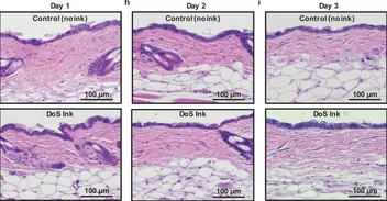
Q. Wang , H. Sheng , Y. Lv , J. Liang , Y. Liu , N. Li , E. Xie , Q. Su , F. Ershad , W. Lan , J. Wang , C. Yu
Advanced Functional Materials · 32(21), 2111228 (2022)
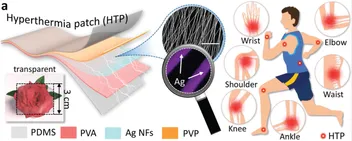
F. Ershad , K. Sim , C. Yu
2022 IEEE 35th International Conference on Micro Electro Mechanical Systems Conference (MEMS) · 39–42 (2022)
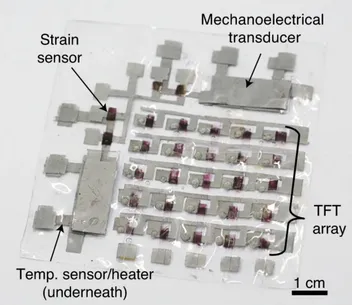
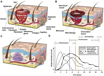
S. Patel , F. Ershad , M. Zhao , R. R. Isseroff , B. Duan , Y. Zhou , Y. Wang , C. Yu
Soft Science · 2(2), 9 (2022)
Prior to University of Houston faculty appointment
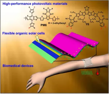
B. Kan , F. Ershad , Z. Rao , C. Yu
Nano Research · 14(9), 2891–2903 (2021)
Prior to University of Houston faculty appointment
K. Sim† , F. Ershad† , Y. Zhang† , P. Yang , H. Shim , Z. Rao , Y. Lu , A. Thukral , A. Elgalad , Y. Xi , B. Tian , D. Taylor , C. Yu
† Equal contribution
Nature Electronics · 3(12), 775–784 (2020)
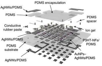
Y.-S. Guan , A. Thukral , S. Zhang , K. Sim , X. Wang , Y. Zhang , F. Ershad , Z. Rao , F. Pan , P. Wang , J. Xiao , C. Yu
Science Advances · 6(38), eabb3656 (2020)
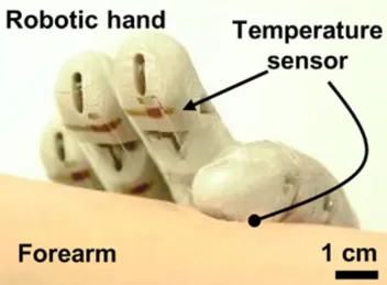
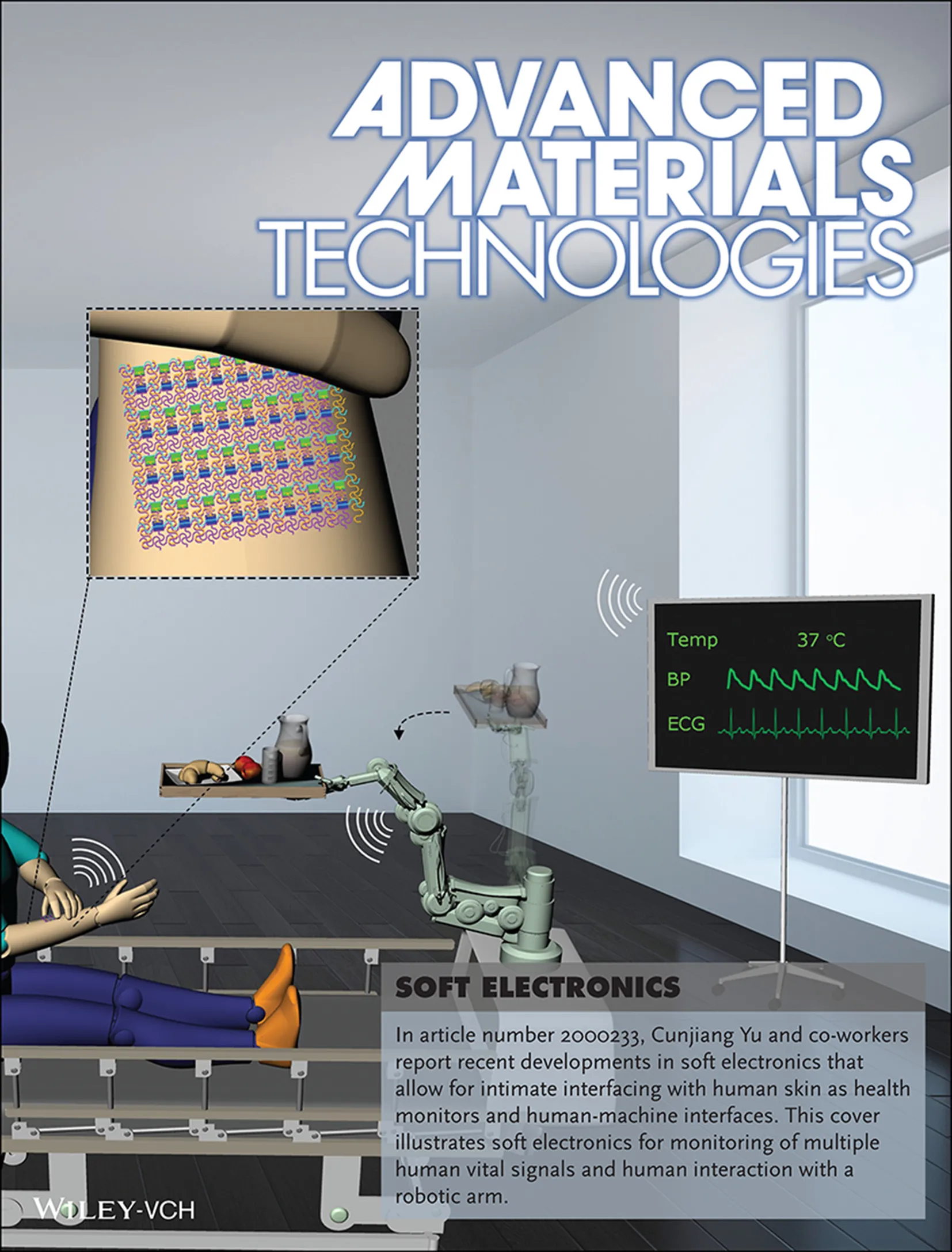
Z. Rao† , F. Ershad† , A. Almasri , L. Gonzalez , X. Wu , C. Yu
† Equal contribution
Advanced Materials Technologies · 5(9), 2000233 (2020)
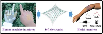
F. Ershad , A. Thukral , J. Yue , P. Comeaux , Y. Lu , H. Shim , K. Sim , N.-I. Kim , Z. Rao , R. Guevara , L. Contreras , F. Peng , Y. Zhang , Y.-S. Guan , P. Yang , X. Wang , P. Wang , X. Wu , C. Yu
Nature Communications · 11(1), 3823 (2020)
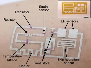
K. Sim , Z. Rao , F. Ershad , C. Yu
Advanced Materials · 32(15), 1902417 (2020)
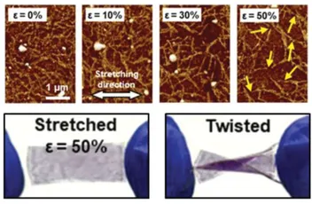
H. Shim , K. Sim , F. Ershad , S. Jang , C. Yu
Journal of Materials Chemistry C · 8(31), 10719–10731 (2020)
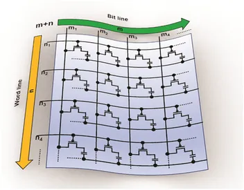
Prior to University of Houston faculty appointment
L. Chang† , Y. Wang† , F. Ershad† , R. Yang , C. Yu , Y. Fan
† Equal contribution
Trends in Biotechnology · 37(11), 1175–1188 (2019)
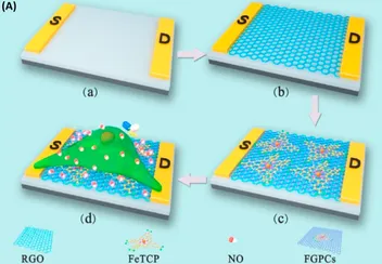
H. Shim , K. Sim , F. Ershad , P. Yang , A. Thukral , Z. Rao , H.-J. Kim , Y. Liu , Xu Wang , G. Gu , L. Gao , Xinran Wang , Y. Chai , C. Yu
Science Advances · 5(10), eaax4961 (2019)
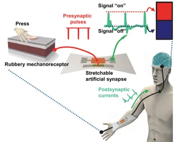
K. Sim , S. Chen , Z. Li , Z. Rao , J. Liu , Y. Lu , S. Jang , F. Ershad , J. Chen , J. Xiao , C. Yu
Nature Electronics · 2(10), 471–479 (2019)
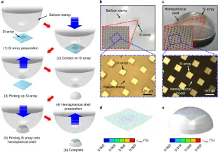
K. Sim , Z. Rao , Z. Zou , F. Ershad , J. Lei , A. Thukral , J. Chen , Q. Huang , J. Xiao , C. Yu
Science Advances · 5(8), eaav9653 (2019)
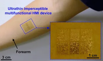
F. Ershad , K. Sim , A. Thukral , Y. S. Zhang , C. Yu
APL Materials · 7(3), 031301 (2019)
Prior to University of Houston faculty appointment
A. Thukral , F. Ershad , N. Enan , Z. Rao , C. Yu
IEEE Nanotechnology Magazine · 12(1), 21–34 (2018)
No publications match those filters.
Select a different research topic, or switch back to “All topics”.